Microscopy has revolutionized the way we see the microscopic world. While traditional light microscopy allows us to observe cell shapes and structures, fluorescent microscopy takes it a step further, revealing the hidden details of cellular life in vibrant colors. At the heart of this technique are fluorescent dyes, molecules that light up specific parts of a cell when exposed to certain wavelengths of light.
What Is Fluorescent Microscopy?
Fluorescent microscopy uses the principle of fluorescence, where certain molecules absorb light at one wavelength (excitation) and emit it at another, longer wavelength (emission). By labeling specific cellular components with fluorescent dyes, scientists can visualize proteins, nucleic acids, lipids, and even entire organelles with astonishing clarity. Read more

How Fluorescent Dyes Work
Fluorescent dyes are like tiny lanterns that attach to specific targets within a cell. When illuminated under a fluorescent microscope, they glow, revealing the location and sometimes the behavior of the molecules they are bound to. Each dye is chosen based on its ability to bind to a particular cellular component and the color it emits.

Some common dyes include:
DAPI : binds to DNA and emits a bright blue light, highlighting the cell nucleus. Read more
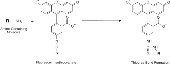
FITC (Fluorescein isothiocyanate) : often used to label antibodies, emitting green fluorescence. Read more
TRITC (Tetramethylrhodamine) : another antibody label, emitting red fluorescence. Read more

Applications in Research
Fluorescent dyes allow researchers to:
Track protein localization in real time.
Study cell division and apoptosis (programmed cell death).
Visualize pathogen-host interactions.
Identify specific cell types in complex tissues.

Tips for Using Fluorescent Dyes
Choose the right dye for your target: Consider specificity, brightness, and photostability.
Minimize photobleaching: Fluorescent dyes can fade with prolonged exposure to light. Use anti-fade mounting media when possible.
Use proper controls: Negative controls help confirm that observed signals are specific to the target molecule.
Conclusion
Fluorescent microscopy dyes have transformed biological research, turning invisible cellular processes into colorful, informative images. From understanding disease mechanisms to discovering new drugs, these dyes are invaluable tools that illuminate the microscopic world like never before.
Our latest content
Check out what's new in our company !